Femoral Fracture
If you are suffering from a Femoral Fracture, please Schedule an appointment with one of our orthopedic specialists as soon as possible.
What is a Femoral Fracture?
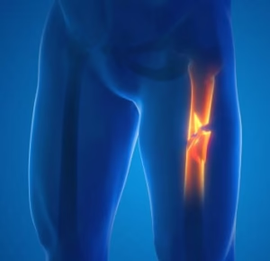 A femoral fracture refers to a break in the femur, the long bone that extends from the hip to the knee. High-energy trauma such as car accidents, falls from significant height, or direct blows often cause the injury, especially in younger individuals. In older adults, weaker bone density from conditions like osteoporosis increases fracture risk even after low-impact falls. Fractures can occur in different regions of the femur, including the head, neck, shaft, or distal end near the knee, and each pattern affects function and stability in different ways.
Common Types of Femoral Shaft Fractures
Transverse fracture: The break forms a straight horizontal line across the femoral shaft.
Oblique fracture: This type of fracture has an angled line across the shaft.
Spiral fracture: The fracture line encircles the shaft like the stripes on a candy cane. A twisting force to the thigh causes this type of fracture.
Comminuted fracture: In this type of fracture, the bone has broken into three or more pieces. In most cases, the number of bone fragments corresponds with the amount of force needed to break the bone.
Open fracture: If a bone breaks in such a way that bone fragments stick out through the skin or a wound penetrates down to the broken bone, the fracture is called an open or compound fracture. Open fractures often involve much more damage to the surrounding muscles, tendons, and ligaments. They have a higher risk for complications—especially infections—and take a longer time to heal.
Examination of a Femoral Fracture
Specialists begin examination of a suspected femoral fracture by assessing the injury history, including mechanism of trauma, pain onset, and ability to bear weight. They inspect the limb for deformity, swelling, bruising, and abnormal positioning while comparing both legs for alignment differences. Palpation helps identify tenderness along the femur, and clinicians evaluate surrounding joints such as the hip and knee for associated injuries. Neurovascular assessment checks distal pulses, skin temperature, sensation, and motor function to rule out blood vessel or nerve damage.
Medical teams confirm diagnosis with imaging studies, most commonly X-rays taken from multiple angles of the femur and adjacent joints. In complex cases, clinicians order CT scans to evaluate fracture patterns or detect subtle breaks. Emergency providers often stabilize the limb with splints or traction during evaluation to prevent further injury and reduce pain. Continuous monitoring ensures early detection of complications such as compartment syndrome or vascular compromise.
Treatment for Femoral Fractures
If you would like to speak to an orthopedic specialist in the DFW metroplex, give us a call at 817-697-4038, or contact us over the web. Tele-medicine appointments are also available.